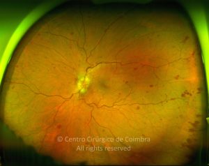
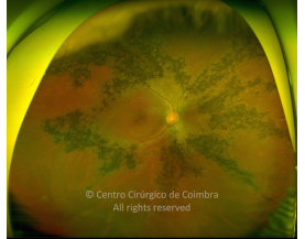
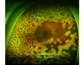
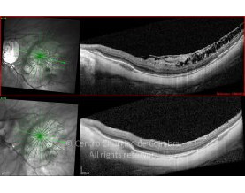
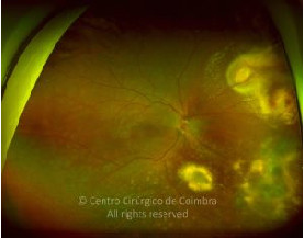
Toxocariasis is an infectious parasitic disease caused by the larvae of the nematodes Toxocara canis and Toxocara cati which are intestinal worms of these hosts. Toxocariasis is a worldwide infection and humans are incidental hosts in which aberrant infection occurs because the parasites cannot completely mature.
The usual mode of infection is eating contaminated soil (geophagia, via hands) or uncooked food. Children who have close contact with dogs (puppies) are at great risk of infection.
The disease is typically unilateral and most often seen in children, though many cases in adults have been reported. Symptoms include decreased visual acuity, but the initial clinical manifestation may be strabismus or leukocoria.
There are three main forms of ocular toxocariasis:
- Peripheral granuloma
- Posterior pole granuloma
- Chronic endophthalmitis
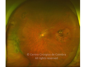
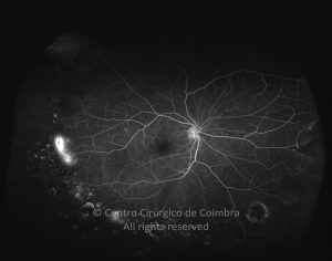
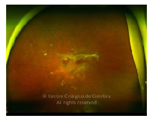
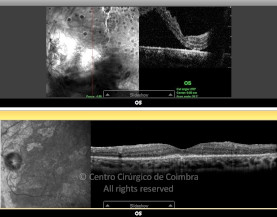
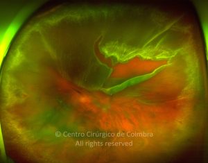
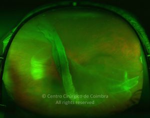
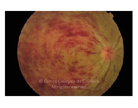
The granuloma is usually yellowish white, 1-2 disc diameter in size, where the larva is encysted. A common finding is a radial fold of the retina extending from the peripheral granuloma to the optic disc. Vitritis may be mild to severe and patients may present with hypopyon.
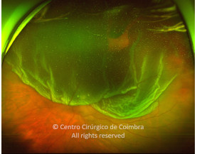
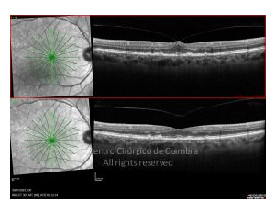
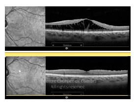
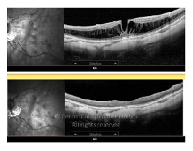
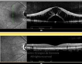
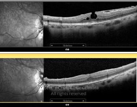
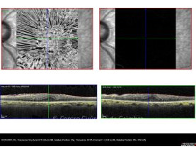
Complications include macular heterotopias, epiretinal membranes, tractional retinal detachment, ciliary body detachment with hypotony, cataract, secondary glaucoma and phthisis bulbi.
Differential diagnosis includes other causes of leukocoria: retinoblastoma, Coats’ disease, retinopathy of prematurity, persistent hyperplastic primary vitreous, familial exudative vitreoretinopathy, toxoplasmosis, pars planitis, and endogenous endophthalmitis.
The diagnosis is based on clinical picture and serologic confirmation of specific IgG and IgM antibodies using ELISA. Other methods are detection of specific anti-toxocara IgG by Western-blot, specific IgE, and intraocular fluids PCR analysis. Patients with visceral larva migrans have leukocytosis and hypereosinophilia, while in patients with ocular toxocariasis they are usually absent.