• Home
  • Who We Are
  • Atlas
  • Dictionary
  • Videos
  • Innovation
  • Blog
  • Contacts
  • English
    • Portuguese (Portugal)
    • French
    • Spanish
  • Home
  • About
  • Atlas
    • Dictionary
  • Videos
  • Blog
  • Innovation
  • Contacts
  • English
    • Portuguese (Portugal)
    • French
    • Spanish
RETINAL VASCULAR DISEASES

Central Retinal Vein Occlusion - Clinical Case 5

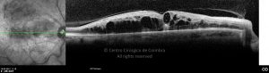
OCT horizontal line-scan demonstrates severe macular edema in a 62-years-old male patient with a central retinal vein occlusion. Visual acuity: 20/200 RE; 20/40 LE.

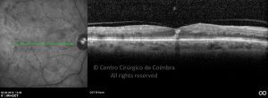
OCT line-scan 10 days after an intraocular injection of dexamethasone implant shows reduction of macular edema, in same case.

Evaluations

1st Evaluation - 15 03 2012

Fluorescein Angiography

  • o59-ti07-2159491-20120315-046
  • o60-ti07-2159491-20120315-047
  • o61-ti07-2159491-20120315-048
  • o62-ti07-2159491-20120315-050
  • o63-ti07-2159491-20120315-051
  • o64-ti07-2159491-20120315-052
  • o65-ti07-2159491-20120315-053
  • o66-ti07-2159491-20120315-054
  • o67-ti07-2159491-20120315-055
  • o68-ti07-2159491-20120315-056
  • o69-ti07-2159491-20120315-057
  • o70-ti07-2159491-20120315-058
  • o71-ti07-2159491-20120315-992
  • o72-ti07-2159491-20120315-993
  • o73-ti07-2159491-20120315-994
  • o74-ti07-2159491-20120315-995
  • o75-ti07-2159491-20120315-998

Autofluorescence

  • o10-ti15-2159491-20120315-004
  • o11-ti15-2159491-20120315-001
  • o12-ti15-2159491-20120315-005
  • o13-ti15-2159491-20120315-006

Infrared

  • o14-ti16-2159491-20120315-008
  • o15-ti16-2159491-20120315-007
  • o16-ti16-2159491-20120315-009

Red-free

  • o08-ti01-2159491-20120315-002
  • o09-ti01-2159491-20120315-003

Biometry

  • o58-ti27-2159491-20120315-001

Anterior Segment Photo

  • o01-ti12-2159491-20120315-001

Microperimetry

  • o56-ti14-2159491-20120315-002
  • o57-ti14-2159491-20120315-001

OCT (B-Scan) longitudinal

  • o19-ti03-2159491-20120315-010
  • o20-ti03-2159491-20120315-011
  • o21-ti03-2159491-20120315-012
  • o22-ti03-2159491-20120315-013
  • o23-ti03-2159491-20120315-014
  • o24-ti03-2159491-20120315-015
  • o25-ti03-2159491-20120315-016
  • o26-ti03-2159491-20120315-017
  • o27-ti03-2159491-20120315-018
  • o28-ti03-2159491-20120315-019
  • o29-ti03-2159491-20120315-020
  • o30-ti03-2159491-20120315-021
  • o31-ti03-2159491-20120315-022
  • o32-ti03-2159491-20120315-023
  • o33-ti03-2159491-20120315-024
  • o34-ti03-2159491-20120315-025
  • o35-ti03-2159491-20120315-026
  • o36-ti03-2159491-20120315-027
  • o37-ti03-2159491-20120315-028
  • o38-ti03-2159491-20120315-029
  • o39-ti03-2159491-20120315-030
  • o40-ti03-2159491-20120315-031
  • o41-ti03-2159491-20120315-032
  • o42-ti03-2159491-20120315-033
  • o43-ti03-2159491-20120315-034
  • o44-ti03-2159491-20120315-035
  • o45-ti03-2159491-20120315-036
  • o46-ti03-2159491-20120315-037
  • o47-ti03-2159491-20120315-038
  • o48-ti03-2159491-20120315-039
  • o49-ti03-2159491-20120315-040
  • o50-ti03-2159491-20120315-041
  • o51-ti03-2159491-20120315-042
  • o52-ti03-2159491-20120315-043
  • o53-ti03-2159491-20120315-044
  • o54-ti03-2159491-20120315-045

Colour Retinography

  • o02-ti02-2159491-20120315-997
  • o04-ti02-2159491-20120315-999
  • o05-ti02-2159491-20120315-001
  • o06-ti02-2159491-20120315-002
  • o07-ti02-2159491-20120315-003
  • o03-ti02-2159491-20120315-996

Tonometry

  • o55-ti21-2159491-20120315-001

2nd Evaluation - 30 03 2012

Autofluorescence

  • o03-ti15-2159491-20120330-002
  • o05-ti15-2159491-20120330-004

Infrared

  • o04-ti16-2159491-20120330-003

Red-free

  • o02-ti01-2159491-20120330-001

OCT (B-Scan) longitudinal

  • o06-ti03-2159491-20120330-005
  • o07-ti03-2159491-20120330-006
  • o08-ti03-2159491-20120330-007
  • o09-ti03-2159491-20120330-008
  • o10-ti03-2159491-20120330-009
  • o11-ti03-2159491-20120330-010

Colour Retinography

  • o01-ti02-2159491-20120330-001

3rd Evaluation - postoperative - 20 04 2012

Fluorescein Angiography

  • o12-ti07-2159491-20120420-008
  • o13-ti07-2159491-20120420-009
  • o14-ti07-2159491-20120420-010
  • o15-ti07-2159491-20120420-011
  • o16-ti07-2159491-20120420-012
  • o17-ti07-2159491-20120420-013

Autofluorescence

  • o10-ti15-2159491-20120420-002

Infrared

  • o11-ti16-2159491-20120420-003

Red-free

  • o07-ti01-2159491-20120420-001

Anterior Segment Photo

  • o01-ti12-2159491-20120420-001

OCT (B-Scan) longitudinal

  • o21-ti03-2159491-20120420-004
  • o22-ti03-2159491-20120420-005
  • o23-ti03-2159491-20120420-006
  • o24-ti03-2159491-20120420-007

Colour Retinography

Ultra-widefield photograph four days after vitrectomy, cataract surgery and endolaser. Note the spatial distribution of laser spots.

  • o03-ti02-2159491-20120420-998
  • o06-ti02-2159491-20120420-001
  • o08-ti02-2159491-20120420-996
  • o18-ti02-2159491-20120420-994
  • o04-ti02-2159491-20120420-997
  • o05-ti02-2159491-20120420-999
  • o09-ti02-2159491-20120420-995
  • o19-ti02-2159491-20120420-993

4th Evaluation - 14 02 2019

Autofluorescence

  • 33-2159491-20190214-002-autof

Infrared

  • 11-2159491-20190214-003-IR

Red-free

  • 11-2159491-20190214-004-red free

OCT (B-Scan) longitudinal

  • 11-2159491-20190214-001
  • 11-2159491-20190214-002

Colour Retinography

VA: 20/400 RE. Focal hemorrhage in the superior macular arcade.

  • 33-2159491-20190214-001

5th Evaluation - 07 11 2019

Autofluorescence

  • 33-2159491-20191107-002-autof

Infrared

  • 11-2159491-20191107-003-IR

Colour Retinography

  • 33-2159491-20191107-001

OCT (B-Scan) longitudinal

Chronic cystoid macular edema in the RE.

  • 11-2159491-20191107-001
  • 11-2159491-20191107-002


  • Rua Dr. Manuel Campos Pinheiro, 51 Espadaneira - S. Martinho do Bispo
    3045-089 Coimbra, Portugal
    (+351) 239 802 700
    www.ccci.pt
    atlasrleye@ccci.pt

  • UPS 07/02.00 (Licença de Funcionamento) Certidão Registo E106499

    Imagiologia:LIC-103/2021 (Licença de Funcionamento)

  • Política de Privacidade

    Política de Cookies


Centro Cirúrgico de Coimbra. Todos os direitos reservados.