• Home
  • Who We Are
  • Atlas
  • Dictionary
  • Videos
  • Innovation
  • Blog
  • Contacts
  • English
    • Portuguese (Portugal)
    • French
    • Spanish
  • Home
  • About
  • Atlas
    • Dictionary
  • Videos
  • Blog
  • Innovation
  • Contacts
  • English
    • Portuguese (Portugal)
    • French
    • Spanish
DIABETIC RETINOPATHY

NON-PROLIFERATIVE DIABETIC RETINOPATHY

Diabetes is a worldwide epidemic disease. In recent decades it has progressed from a disease affecting people primarily in developed countries, to a global phenomenon. Today diabetic retinopathy is a major cause of blindness in adult patients.

The eye is one of the target organs of this multisystemic disease. Diabetic retinopathy is nothing more than a compromise of the retinal microcirculation. The chronic exposure to hyperglycemia determines a number of biochemical and consequently histological changes. Initially, selective loss of pericytes, basement membrane thickening, as well as a diverse number of hematologic abnormalities, generate capillary occlusion, microaneurysm formation, dilation and beading of retinal veins as well as retinal ischemia.

A compromise of the endothelial barrier function allows vascular leakage, originating retinal edema and deposition of hard exsudates. Areas of retinal ischemia generate vasoproliferative factors (mainly vascular endothelium growth factor – VEGF) that later stimulate the growth of neovessels.

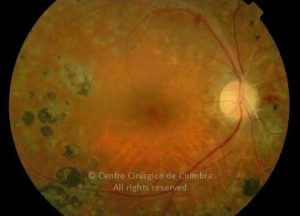
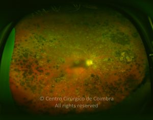
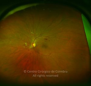
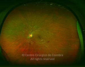
According to fundoscopic findings, diabetic retinopathy may be divided in non-proliferative and proliferative diabetic retinopathy, based on the presence of neovascularization. The most commonly used classification scale is the International Clinical Diabetic Retinopathy Disease Severity Scale:

0
Clinical Cases

No apparent retinopathy: no abnormalities

  • Mild Non-proliferative Diabetic Retinopathy (NPDR): microaneurysms only
  • Moderate NPDR: more than just microaneurysms but less than severe NPDR
  • Severe NPDR:

Any of the following:

  • More than 20 intraretinal hemorrhages in each of 4 quadrants
  • Definite venous beading in 2 or more quadrants
  • Prominent intraretinal microvascular abnormalities (IRMA) in at least one quadrant

And no signs of proliferative retinopathy

  • Proliferative Diabetic retinopathy

One or more of the following:

  • Neovascularization
  • Vitreous/Preretinal hemorrhage

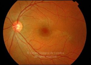
Vision loss in diabetic patients can have several causes: macular oedema, macular ischemia and sequelae of neovascularization, due to induced ischemia.

Medical management plays a primordial role in PDR. Medical treatment plays a key role in PDR. Glycemic control (HbA1c – should be between 5.5-6.5% according to the American Academy of Endocrinology) and the blood pressure control is essential for a good result.

Panretinal photocoagulation (PRP) is the gold standard treatment for patients with severe NPDR and PDR. Laser photocoagulation aims to destroy the ischemic areas of the retina and to reduce the production of vasoproliferative factors It is however associated with significant side effects such as: visual field loss, dischromatopsia and decreased night vision. It may also exacerbate a macular edema. Therefore, PRP should be judiciously used and macular edema addressed prior to PRP (see below).

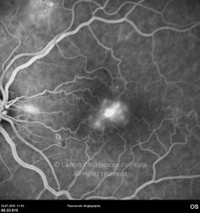
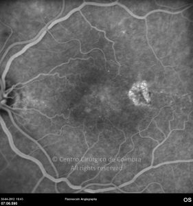
Diabetic macular edema is a serious complication of the increased vascular permeability. It can be characterized clinically as focal, multifocal and diffuse The concept of clinically significant macular edema (CSME) was first introduced in the Early Treatment Diabetic Retinopathy Study (ETDRS).

CSME was defined as retinal edema located at or within 500 µm of the center of the macula, hard exsudates at or within 500 µm of the center of the macula if associated with thickening of the adjacent retina or a zone of thickening larger than 1 disc area if located within 1 disc diameter of the center of the macula.

ETDRS trial found a better visual outcome for patients with CSME submitted to focal laser (50% of risk reduction in doubling the visual angle). Anti-VEGF agents have also been progressively used to treat CSME CMSE can be exacerbated by PRP or cataract surgery and therefore, its treatment should take precedence.

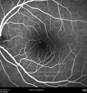
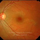
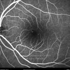
Ischemic maculopathy is characterized by an increase in diameter of the foveal avascular zone as well as an irregularity of its limits. Fluorescein angiography allows the definitive diagnosis. There is no proven treatment for ischemic maculopathy.

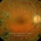
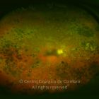
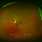
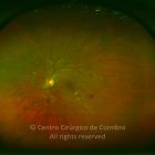
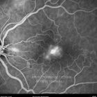
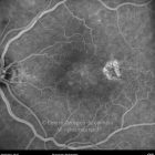
Clinical Cases

Clinical Case 1

6

65

0

Clinical Case 2

8

101

0

Clinical Case 3

8

95

0

Clinical Case 4

9

70

0


  • Rua Dr. Manuel Campos Pinheiro, 51 Espadaneira - S. Martinho do Bispo
    3045-089 Coimbra, Portugal
    (+351) 239 802 700
    www.ccci.pt
    atlasrleye@ccci.pt

  • UPS 07/02.00 (Licença de Funcionamento) Certidão Registo E106499

    Imagiologia:LIC-103/2021 (Licença de Funcionamento)

  • Política de Privacidade

    Política de Cookies


Centro Cirúrgico de Coimbra. Todos os direitos reservados.