OPTIC NERVE DISEASES
Optic Disc Pits and Associated Serous Macular Detachments
Clinical Case 03
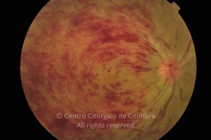
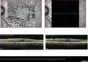
Neurosensorial detachment and a bilaminar structure "schisis-like" between external and internal retinal layers associated to an optic disc pit, in a 10-years-old patient
Evaluations
1st evaluation
46
0
13 07 2011
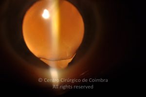
- Anterior Segment Photo
13 07 2011
- Autofluorescence
13 07 2011
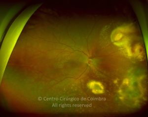
- Colour Retinography
13 07 2011
- Infrared
13 07 2011
- Microperimetry
13 07 2011
- OCT B-Scan (Longitudinal)
13 07 2011
- Red-free
13 07 2011
- RNFL - Thickness Image
2nd evaluation
29
0
22 08 2011
- Autofluorescence
22 08 2011
- Colour Retinography
22 08 2011
- Infrared
13 07 2011
- Microperimetry
22 08 2011
- OCT B-Scan (Longitudinal)
22 08 2011
- Red-free
29 08 2011
- Tonometry
3rd evaluation - postoperative
9
0
4th evaluation
13
0
14 10 2011
- Autofluorescence
12 09 2011
- Colour Retinography
14 10 2011
- OCT B-Scan (Longitudinal)
14 10 2011
- Red-free
5th evaluation
37
0
16 03 2012
- Autofluorescence
16 03 2012
- Colour Retinography
16 03 2012
- Infrared
16 03 2012
- OCT B-Scan (Longitudinal)
16 03 2012
- Red-free
6th evaluation
55
0
07 05 2012
- Anterior Segment Photo
07 05 2012
- Autofluorescence
07 05 2012
- Colour Retinography
07 05 2012
- Infrared
07 05 2012
- OCT B-Scan (Longitudinal)
07 05 2012
- Red-free
7th evaluation
13
0
19 07 2013
- Autofluorescence
19 07 2013
- Colour Retinography
19 07 2013
- Infrared
19 07 2013
- OCT B-Scan (Longitudinal)
19 07 2013
- Red-free
8th evaluation
8
0
29 08 2014
- Colour Retinography
29 08 2014
- Infrared
29 08 2014
- OCT B-Scan (Longitudinal)
29 08 2014
- Red-free
9th evaluation
8
0
30 03 2016
- Colour Retinography
30 03 2016
- Infrared
30 03 2016
- OCT B-Scan (Longitudinal)
30 03 2016
- Red-free
10th evaluation
9
0