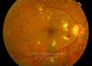
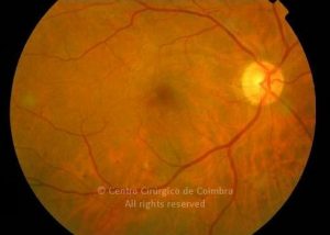
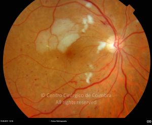
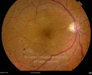
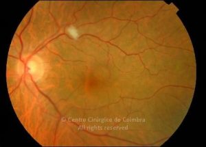
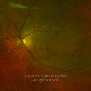
Uma mancha algodonosa é uma lesão branca, na superfície da retina, e de limites mal definidos.
Estas lesões correspondem a áreas focais de não-perfusão dos capilares retinianos, resultando numa interrupção isquémica do transporte axonal e edema na camada de fibras nervosas da retina.
Anomalias Associadas com Manchas Algodonosas no Fundo Ocular:
- Síndroma de imunodeficiência adquirida
- Anemia aguda
- Pancreatite aguda
- Síndroma do arco aórtico
- Doença valvular cardíaca
- Aterosclerose carotídea
- Oclusão da veia central e de ramo da retina
- Doenças vasculares do colagénio
- Retinopatia diabética
- Disproteinémias
- Abuso de drogas intravenosas
- Leptospirose
- Leucemia
- Carcinoma metastático
- Oncocercose
- Edema do disco ótico
- Papilite
- Oclusão de ramo da artéria central da retina
- Retinopatia da radiação
- Retinopatia da “Rocky Mountain spotted fever”
- Septicémia
- Anemia grave
- Hipertensão arterial sistémica
- Administração sistémica interferão alfa
- Traumatismo


