RETINAL DYSTROPHIES AND SYNDROMES
Wyburn-Mason Syndrome
Clinical Case 01
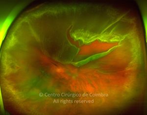
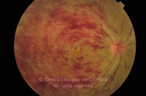
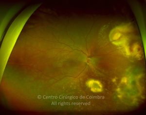
Fundus photograph of Wiburn-Mason Syndrome in a 40-years-old male patient. Note the tortuous retinal artery
Evaluations
1st evaluation
12
0
17 11 2008
- Autofluorescence
17 11 2008
- Colour Retinography
17 11 2008
- Fluorescein Angiography
17 11 2008
- Infrared
17 11 2008
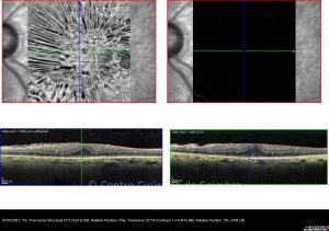
- OCT B-Scan (Longitudinal)
17 11 2008
- Red-free
2nd evaluation - postoperative
13
0
16 01 2009
- Anterior Segment Photo
26 01 2009
- Autofluorescence
19 01 2009
- Colour Retinography
04 03 2009
- Fluorescein Angiography
26 01 2009
- Infrared
26 01 2009
- OCT B-Scan (Longitudinal)
26 01 2009
- Red-free
3rd evaluation
13
0
19 10 2009
- Anterior Segment Photo
19 10 2009
- Autofluorescence
19 10 2009
- Colour Retinography
19 10 2009
- Fluorescein Angiography
19 10 2009
- Infrared
19 10 2009
- Red-free
4th evaluation
5
0
30 09 2010
- Autofluorescence
30 09 2010
- Colour Retinography
30 09 2010
- Infrared
30 09 2010
- Red-free
5th evaluation
44
0
04 05 2011
- Autofluorescence
04 05 2011
- Colour Retinography
04 05 2011
- Fluorescein Angiography
04 05 2011
- Infrared
04 05 2011
- OCT B-Scan (Longitudinal)
04 05 2011
- Red-free
04 05 2011
- Tonometry
6th evaluation
8
0
23 11 2011
- Autofluorescence
23 11 2011
- Colour Retinography
23 11 2011
- Infrared
23 11 2011
- Perimetry
23 11 2011
- Red-free
7th evaluation
21
0
12 06 2012
- Anterior Segment Photo
12 06 2012
- Autofluorescence
12 06 2012
- Colour Retinography
12 06 2012
- Infrared
12 06 2012
- Nervous Fiber Analysis
12 06 2012
- OCT B-Scan (Longitudinal)
12 06 2012
- Red-free
8th evaluation
6
0