MACULAR DISEASES
Hypotonic Maculopathy
Clinical Case 01
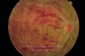
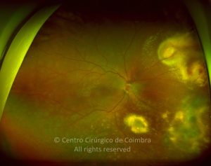
Fundus photograph of hypotonic eye, 2 months after traumatic injury. Note the chorioretinal folds in the posterior pole, papilloedema and vascular tortuosity. Visual acuity: 20/100 LE
Evaluations
1st evaluation
27
0
08 06 2011
- Anterior Segment Photo
21 06 2011
- Autofluorescence
21 06 2011
- Colour Retinography
21 06 2011
- Fluorescein Angiography
21 06 2011
- Infrared
21 06 2011
- Microperimetry
21 06 2011
- OCT B-Scan (Longitudinal)
21 06 2011
- Red-free
21 06 2011
- Tonometry
2nd evaluation
5
0
3rd evaluation
10
0
12 09 2011
- Autofluorescence
12 09 2011
- Colour Retinography
12 09 2011
- Echography
12 09 2011
- OCT B-Scan (Longitudinal)
12 09 2011
- Red-free
4th evaluation
11
0
24 10 2011
- Anterior Segment Photo
24 10 2011
- Autofluorescence
24 10 2011
- Colour Retinography
24 10 2011
- Infrared
24 10 2011
- OCT B-Scan (Longitudinal)
24 10 2011
- Red-free
24 10 2011
- Tonometry
5th evaluation
13
0
06 01 2012
- Anterior Segment Photo
06 01 2012
- Autofluorescence
06 01 2012
- Colour Retinography
06 01 2012
- Infrared
06 01 2012
- OCT B-Scan (Longitudinal)
06 01 2012
- Red-free
06 01 2012
- Tonometry
6th evaluation - postoperative
7
0
17 01 2012
- Anterior Segment Photo
17 01 2012
- Autofluorescence
17 01 2012
- Colour Retinography
17 01 2012
- Infrared
17 01 2012
- OCT B-Scan (Longitudinal)
17 01 2012
- Red-free
7th evaluation
7
0
20 01 2012
- Anterior Segment Photo
20 01 2012
- Autofluorescence
20 01 2012
- Colour Retinography
20 01 2012
- OCT B-Scan (Longitudinal)
20 01 2012
- Red-free
8th evaluation
12
0