About
Atlas
Videos
Present
EN
-
PT
-
ES
-
FR
About
Atlas
Videos
Present
EN
-
PT
-
ES
-
FR
Home
Atlas
RETINAL AND CHOROIDAL TUMORS
Choroidal Nevus
Clinical Case 01
Clinical Case 01
Filter
Filter
Chapters
All
AGE-RELATED MACULAR DEGENERATION
ANOTHER CLINICAL CASES
CHORIORETINAL INFLAMMATORY DISEASES - Infectious Causes
CHORIORETINAL INFLAMMATORY DISEASES - Noninfectious Causes
CONGENITAL AND PEDIATRIC RETINAL DISEASES
DIABETIC RETINOPATHY
MACULAR DISEASES
OPTIC NERVE DISEASES
PERIPHERAL RETINAL DISEASES
RETINAL AND CHOROIDAL TUMORS
RETINAL DYSTROPHIES AND SYNDROMES
RETINAL VASCULAR DISEASES
TRAUMATIC AND TOXIC RETINOPATHIES
VITREOUS DISEASES
Diseases
Clinical Cases
Clinical Cases
Age (range)
Intervention Types
All
Angiography with Green Indocyanine
Anterior Segment Photo
Autofluorescence
Biometry
Colour Retinography
Computed Radiography
Computed Tomography
Contrast sensitivity / Colour sensitivity
Corneal Topography
Echography
ERG, multifocal
ERG, pattern
Fluorescein Angiography
Infrared
Magnetic Ressonance
Microperimetry
Nervous Fiber Analysis
OCT B-Scan (Longitudinal)
OCT C-Scan (Coronal)
OCT, Cornea
Perimetry
Red-free
Refraction
RNFL - Nervous Fiber Analysis
RNFL - Thickness Image
Specular Microscopy
Surgical Video
Tonometry
With images
With videos
New
Clean
Apply
RETINAL AND CHOROIDAL TUMORS
Choroidal Nevus
Clinical Case 01
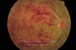
Fundus photograph in a 55-years-old female patient showing a choroidal nevus. Note the drusen on the surface of the tumour
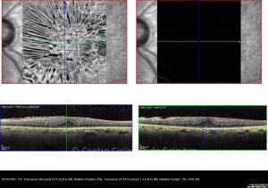
OCT horizontal line-scan of choroidal nevus, in same case
Evaluations
1st evaluation
28
0
05 01 2012
- Autofluorescence
05 01 2012
- Colour Retinography
05 01 2012
- Infrared
05 01 2012
- OCT B-Scan (Longitudinal)
05 01 2012
- Red-free