OPTIC NERVE DISEASES
Optic Disc Drusen
Clinical Case 03
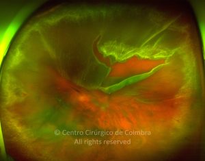
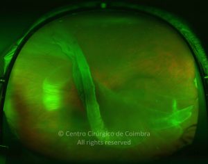
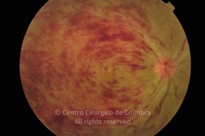
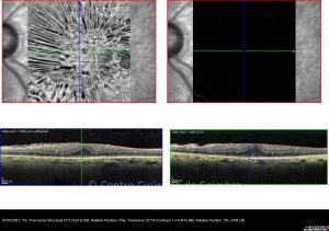
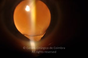
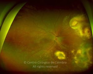
Fundus photograph in a 67-years-old male patient with optic disc drusen, cataract and a macular epiretinal membrane. Visual acuity: 20/40 RE; 20/100 LE
Evaluations
1st evaluation
49
0
11 01 2012
- Anterior Segment Photo
11 01 2012
- Autofluorescence
11 01 2012
- Biometry
11 01 2012
- Colour Retinography
16 01 2012
- Echography
11 01 2012
- Fluorescein Angiography
11 01 2012
- Infrared
11 01 2012
- Microperimetry
11 01 2012
- Nervous Fiber Analysis
11 01 2012
- OCT B-Scan (Longitudinal)
11 01 2012
- Perimetry
11 01 2012
- Red-free
11 01 2012
- RNFL - Nervous Fiber Analysis
11 01 2012
- RNFL - Thickness Image
11 01 2012
- Tonometry
2nd evaluation - postoperative
10
0
17 01 2012
- Anterior Segment Photo
17 01 2012
- Autofluorescence
17 01 2012
- Colour Retinography
17 01 2012
- Infrared
17 01 2012
- OCT B-Scan (Longitudinal)
17 01 2012
- Red-free
3rd evaluation
8
0
25 01 2012
- Autofluorescence
21 02 2012
- Colour Retinography
25 01 2012
- OCT B-Scan (Longitudinal)
25 01 2012
- Tonometry
4th evaluation
10
0
02 03 2012
- Anterior Segment Photo
02 03 2012
- Autofluorescence
02 03 2012
- Colour Retinography
02 03 2012
- Infrared
02 03 2012
- OCT B-Scan (Longitudinal)
02 03 2012
- Red-free
4th evaluation - postoperative
10
0
02 03 2012
- Anterior Segment Photo
02 03 2012
- Autofluorescence
02 03 2012
- Colour Retinography
02 03 2012
- Infrared
02 03 2012
- OCT B-Scan (Longitudinal)
02 03 2012
- Red-free
5th evaluation
8
0
12 03 2012
- Autofluorescence
12 03 2012
- Colour Retinography
12 03 2012
- Infrared
12 03 2012
- OCT B-Scan (Longitudinal)
12 03 2012
- Tonometry
6th evaluation
17
0